
MSK Ultrasound Course: Diagnosing Musculoskeletal Injuries with Precision
Musculoskeletal injuries are common in sports medicine, orthopedics, rheumatology, rehabilitation, and emergency care. From shoulder pain and tendon injuries to […]
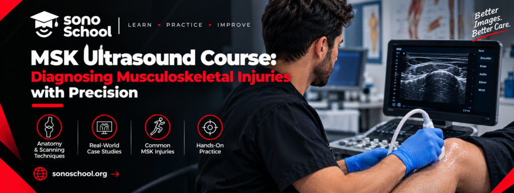
Musculoskeletal injuries are common in sports medicine, orthopedics, rheumatology, rehabilitation, and emergency care. From shoulder pain and tendon injuries to ligament problems and soft tissue swelling, clinicians need reliable methods to assess musculoskeletal structures and make informed treatment decisions.
Musculoskeletal ultrasound (MSK ultrasound) is a valuable imaging technique that allows trained clinicians to examine tendons, muscles, ligaments, joints, bursae, and selected peripheral nerves in real time. Unlike imaging methods that require a separate appointment, ultrasound can be performed at the point of care when appropriate equipment and trained professionals are available.
However, accurate interpretation requires more than operating an ultrasound machine. Clinicians must understand anatomy, use systematic scanning techniques, recognize normal and abnormal findings, and correlate imaging results with the patient’s symptoms and physical examination.
An MSK ultrasound course can help healthcare professionals develop these skills through structured education and hands-on practice.
In this guide, we explain what musculoskeletal ultrasound training involves, which injuries it can help assess, what to look for when choosing a course, and how to select the right training program for your professional goals.
What Is an MSK Ultrasound Course?
An MSK ultrasound course is a structured training program that teaches healthcare professionals how to examine musculoskeletal structures using ultrasound.
The curriculum typically covers ultrasound physics, probe selection, image optimization, musculoskeletal anatomy, scanning techniques, and the interpretation of selected abnormalities.
Depending on the course, participants may learn to assess:
- Tendons and tendon injuries.
- Muscles and soft tissue abnormalities.
- Ligaments and selected joint structures.
- Joint effusions and bursae.
- Shoulder, elbow, wrist, hand, hip, knee, ankle, and foot anatomy.
- Selected peripheral nerves.
- Common findings associated with musculoskeletal pain and injury.
The best course combines theoretical instruction with supervised scanning practice. This helps participants understand how to obtain appropriate images and recognize the limitations of ultrasound examinations.
For physicians who want to develop practical MSK ultrasound skills, SonoSchool’s Musculoskeletal Ultrasound Course offers a one-day, hands-on introduction to scanning major joints, tendons, and soft tissues.
Why Is Musculoskeletal Ultrasound Important in Clinical Practice?
Musculoskeletal ultrasound provides dynamic, real-time imaging of accessible soft tissue structures. It can complement clinical examination and help answer focused questions about pain, swelling, injury, or selected joint abnormalities.
1. Assess Tendon Injuries
Ultrasound can help evaluate tendons such as the Achilles tendon, rotator cuff tendons, and patellar tendon.
Depending on the location and clinical question, findings may include tendon thickening, altered echotexture, fluid around a tendon, or evidence of a tear.
Dynamic assessment can also help clinicians examine how a structure behaves during movement.
2. Examine Muscles and Soft Tissues
Ultrasound can help assess selected muscle injuries, localized swelling, superficial soft tissue abnormalities, and fluid collections.
For sports medicine and rehabilitation professionals, this can provide useful information when evaluating an injury alongside the patient’s history and physical examination.
3. Evaluate Joint Effusions and Bursae
Ultrasound can identify fluid in accessible joints and bursae. For example, it may help assess a knee effusion or evaluate the subacromial-subdeltoid bursa in a patient with shoulder pain.
The presence of fluid does not automatically establish the underlying diagnosis. Clinical correlation remains essential.
4. Support Dynamic Assessment
One important advantage of ultrasound is the ability to assess selected structures during movement.
Dynamic scanning may help demonstrate tendon movement, certain instability patterns, or abnormalities that are less apparent during a static examination.
The examination must be performed using appropriate techniques, and the findings should be interpreted within the clinical context.
For further reference, consult the American Institute of Ultrasound in Medicine (AIUM) Musculoskeletal Practice Parameters, which outline principles for performing and documenting musculoskeletal ultrasound examinations.
What Will You Learn in an MSK Ultrasound Training Course?
A comprehensive musculoskeletal ultrasound course should combine foundational knowledge with practical scanning experience.
1. Ultrasound Physics and Image Optimization
Participants learn how ultrasound images are produced and how machine settings affect image quality.
Important topics include probe selection, depth, gain, focus, image orientation, and common ultrasound artifacts.
Understanding artifacts is particularly important in MSK ultrasound because anisotropy can make normal tendons appear artificially hypoechoic when the ultrasound beam is not appropriately aligned with the tendon fibers.
2. Shoulder Ultrasound
Shoulder scanning is an important part of musculoskeletal ultrasound training.
Depending on the curriculum, participants may learn to examine the rotator cuff tendons, long head of the biceps tendon, subacromial-subdeltoid bursa, and selected surrounding structures.
These techniques can help clinicians investigate shoulder pain and selected tendon or soft tissue abnormalities.
3. Elbow, Wrist, and Hand Ultrasound
Training may include systematic assessment of the elbow’s anterior, medial, lateral, and posterior regions.
Wrist and hand scanning can cover tendons, selected joints, the carpal tunnel region, and superficial soft tissue structures.
These applications may be useful in evaluating selected tendon disorders, localized swelling, and symptoms that require further assessment.
4. Hip and Knee Ultrasound
Hip ultrasound training may include selected joint and periarticular structures, effusion assessment, and soft tissue evaluation.
Knee scanning can cover the suprapatellar recess, quadriceps and patellar tendons, selected ligaments, and the region associated with a Baker’s cyst.
The appropriate examination depends on the patient’s symptoms and the clinical question being investigated.
5. Ankle and Foot Ultrasound
Ankle and foot applications may include the Achilles tendon, ankle tendons, selected ligaments, plantar fascia, and other accessible soft tissue structures.
These skills are relevant to clinicians assessing sports injuries, overuse conditions, and selected causes of foot and ankle pain.
SonoSchool’s MSK Ultrasound Course includes training across the shoulder, elbow, wrist, hand, hip, knee, ankle, and foot, with supervised practical scanning and clinical integration.
Who Should Enroll in an MSK Ultrasound Course?
Musculoskeletal ultrasound training may benefit several healthcare professionals, depending on their scope of practice and clinical responsibilities.
| Specialty | Potential applications |
| Orthopedics | Selected tendon, ligament, joint, and soft tissue assessments |
| Sports medicine | Evaluation of selected sports-related injuries and tendon abnormalities |
| Rheumatology | Assessment of selected joint, tendon, and inflammatory abnormalities |
| Physical medicine and rehabilitation | Musculoskeletal assessment to support rehabilitation planning |
| Emergency medicine | Focused assessment of selected superficial injuries and soft tissue conditions |
| General practice | Selected musculoskeletal presentations within the clinician’s competence |
An introductory course may be suitable for clinicians who are new to MSK ultrasound. Those seeking more advanced diagnostic or interventional skills should look for additional training appropriate to their specialty.
How to Choose the Best MSK Ultrasound Course
Before you register for an MSK ultrasound course, compare the educational content, practical training, instructors, and learning objectives.
Look for Hands-On Scanning
Musculoskeletal ultrasound is a practical skill. Choose a course that provides supervised scanning opportunities rather than relying entirely on lectures.
Ask how much time is allocated to practical sessions and whether participants receive individual feedback.
Check the Course Curriculum
The curriculum should cover the anatomical regions and applications most relevant to your clinical practice.
If you frequently assess shoulder injuries, for example, make sure the course provides sufficient time for shoulder scanning and interpretation.
Evaluate Faculty and Feedback
Look for instructors with relevant clinical expertise and ultrasound teaching experience.
A good training program explains how to obtain appropriate views, recognize common technical errors, interpret findings, and identify situations in which ultrasound alone is insufficient.
Verify Certification and Accreditation Claims
Before paying, ask what certificate the course provides and whether any CME credits or formal accreditation apply.
A certificate of completion is not automatically equivalent to professional certification or independent clinical credentialing. Verify the awarding organization and the exact requirements before enrolling.
The AIUM Diagnostic MSK Ultrasound Accreditation Resources provide information about requirements and practice parameters for diagnostic musculoskeletal ultrasound accreditation.
Compare Cost, Schedule, and Practical Value
Consider the course fee, location, duration, practical hours, and any additional expenses.
The most affordable course is not necessarily the best choice. Training quality, instructor feedback, and opportunities to develop scanning competency are important factors when comparing programs.
MSK Ultrasound Course at SonoSchool: How to Register
SonoSchool offers a one-day Musculoskeletal Ultrasound Course focused on practical scanning of major joints, tendons, and soft tissues.
The published curriculum includes introductory MSK ultrasound principles, image optimization, anatomical orientation, shoulder and elbow scanning, wrist and hand assessment, hip and knee ultrasound, ankle and foot applications, and clinical integration.
The course is intended for clinicians including emergency medicine physicians, orthopedic physicians, rheumatologists, sports medicine physicians, rehabilitation physicians, general practitioners, and residents.
Before registering, confirm the following:
- Upcoming course dates and training location.
- Current tuition fees and registration requirements.
- The amount of supervised practical scanning.
- Instructor qualifications and assessment arrangements.
- Whether a completion certificate or CME credits are available.
To request current details, visit the SonoSchool MSK Ultrasound Course page or contact the SonoSchool team.
Frequently Asked Questions About MSK Ultrasound Courses
1. What is an MSK ultrasound course?
An MSK ultrasound course teaches healthcare professionals how to examine musculoskeletal structures using ultrasound. Training may include tendons, muscles, ligaments, joints, bursae, selected nerves, and common soft tissue abnormalities.
2. Can beginners enroll in an MSK ultrasound course?
Many introductory courses are suitable for clinicians with limited previous experience. Check whether the program covers ultrasound physics, probe handling, image optimization, anatomy, and supervised practical scanning.
3. What injuries can musculoskeletal ultrasound help assess?
Depending on the examination and the operator’s training, ultrasound can help assess selected tendon tears, tendinopathy, muscle injuries, joint effusions, bursitis, and superficial soft tissue abnormalities. It does not detect every injury, and further imaging may be necessary.
4. How long does an MSK ultrasound course take?
Duration varies by provider and course level. SonoSchool lists a one-day introductory MSK ultrasound course. More advanced competencies generally require additional supervised practice and experience.
5. Is MSK ultrasound better than MRI?
Neither modality is universally better. Ultrasound offers real-time dynamic assessment of accessible structures, while MRI provides a broader view of many deep and complex anatomical regions. The most appropriate investigation depends on the suspected injury, clinical findings, and local protocols.
6. Does an MSK ultrasound course provide certification?
This depends on the provider. Some courses provide a completion certificate, while others may offer CME credits or operate under a particular accreditation framework. Confirm the exact certificate, awarding organization, and eligibility requirements before enrolling.
7. Where can I register for an MSK ultrasound course?
You can review the SonoSchool Musculoskeletal Ultrasound Course and contact the team to request current dates, fees, location, and enrollment information.
Conclusion: Develop Your Musculoskeletal Ultrasound Skills
An MSK ultrasound course can help clinicians develop practical skills for assessing selected musculoskeletal injuries and abnormalities. By combining anatomical knowledge, systematic scanning, and image interpretation, healthcare professionals can learn to use ultrasound more effectively within their clinical practice.
When selecting a course, prioritize supervised hands-on training, relevant anatomical coverage, experienced instructors, and transparent information about certification and course requirements.
SonoSchool offers a one-day introductory musculoskeletal ultrasound course covering major joints, tendons, and soft tissues. Review the curriculum, confirm the current registration details, and determine whether the course matches your professional goals.
Ready to improve your musculoskeletal ultrasound skills? Explore the SonoSchool MSK Ultrasound Course and contact the team to learn more about upcoming training opportunities.
Medical education disclaimer: This article is for educational purposes. Ultrasound findings should be interpreted by appropriately trained clinicians in conjunction with clinical assessment and relevant investigations. Training completion does not automatically establish competence in every examination or procedure.
Recommended learning path
Choose the next step based on whether you want to read more, join a workshop, or explore structured fellowship training.



Ready to build real bedside ultrasound confidence?
Join structured POCUS courses and hands-on workshops designed to move clinicians from theory to confident bedside application.