
From Pocket to Bedside: Building Handheld POCUS Into Your Workflow
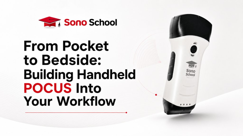
POCUS Workflow Owning a handheld ultrasound probe is not the same as using POCUS well. A handheld ultrasound device can sit in your pocket, on your desk, or…
POCUS Workflow
Owning a handheld ultrasound probe is not the same as using POCUS well.
A handheld ultrasound device can sit in your pocket, on your desk, or inside your department. But unless it becomes part of a clear clinical workflow, it may remain underused, inconsistently used, or used without enough structure.
The real value of handheld POCUS comes when clinicians know when to scan, what question to ask, how to acquire the image, how to interpret the finding, how to document it, and when to escalate beyond a focused bedside scan.
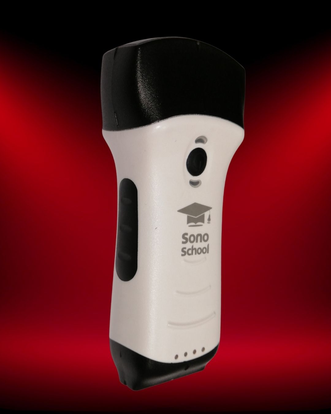
This article is for clinicians and healthcare facilities that already understand the potential of handheld ultrasound, but want to move from “we have a probe” to “we use POCUS consistently and responsibly.”
1. Start With the Clinical Question, Not the Device
The first step in building handheld POCUS into clinical workflow is deciding what questions the device will answer.
POCUS is not “general scanning.” It is focused bedside ultrasound used to support a specific clinical decision. The question should be clear before the probe touches the patient.
Examples of focused clinical questions include:
- Is there evidence of pleural effusion?
- Are there diffuse B-lines suggesting interstitial syndrome?
- Is lung sliding present in this clinical context?
- Is there free fluid in a trauma assessment?
- Is there grossly reduced LV function?
- Is there pericardial effusion?
- Can I identify the vessel before vascular access?
- Is the bladder distended?
- Does the procedure site look safe to approach?
This is different from asking, “What can I find if I scan?”
A handheld ultrasound device should support focused decision-making, not random scanning. Without a defined question, clinicians are more likely to over-scan, over-interpret, or misapply findings outside their competence.
Workflow rule: Before scanning, ask: “What clinical decision will this scan help me make?”
2. Define the Core Applications for Your Team
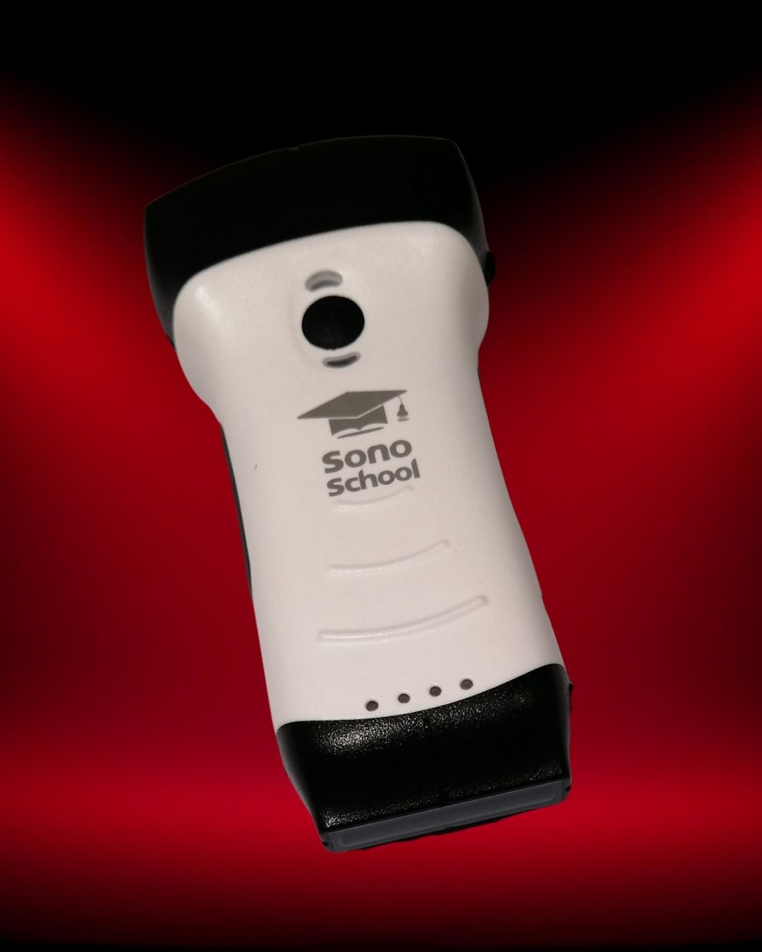
A handheld ultrasound probe can support many clinical applications, but most clinicians do not need to start with everything at once.
A safer and more practical approach is to define a focused application set based on your specialty and workflow.
For example:
- Emergency medicine: lung ultrasound, FAST/eFAST, cardiac POCUS, vascular access, procedures.
- Critical care: lung ultrasound, focused cardiac assessment, vascular access, fluid tolerance assessment, procedural guidance.
- Anesthesia: vascular access, nerve blocks, airway-related assessment where appropriate, procedural guidance.
- Internal medicine: lung, cardiac, abdomen, bladder, volume assessment, ward reassessment.
- General practice or clinics: focused abdomen, bladder, soft tissue, vascular access, screening questions where appropriate.
- Surgery: FAST/eFAST, procedural planning, soft tissue, vascular access, focused abdominal assessment.
Once the application set is defined, the device decision becomes clearer. You can evaluate whether the probe configuration, presets, depth, resolution, software, and export workflow fit those applications.
3. Build a Simple “When to Scan” Pathway
Handheld ultrasound becomes more consistent when clinicians know when it should be used.
This does not require a complex policy at the beginning. It can start with a simple department-level or clinic-level pathway.
Examples:
- Patients with acute dyspnea: consider focused lung ultrasound where trained staff are available.
- Undifferentiated shock: consider focused cardiac, lung, and abdominal views as part of resuscitation assessment.
- Difficult vascular access: use ultrasound before repeated blind attempts.
- Suspected urinary retention: consider bladder assessment where clinically appropriate.
- Procedural planning: assess anatomy before selected bedside procedures.
- High-risk reassessment: repeat focused scanning when clinical status changes.
The goal is not to force ultrasound into every case. The goal is to make handheld POCUS available in situations where it can answer a focused question and influence the next decision.
Workflow rule: Define situations where handheld ultrasound should be considered — and situations where formal imaging or expert review should not be delayed.
4. Create a Standard Image Acquisition Approach
Having a handheld ultrasound device does not mean everyone will scan the same way.
One clinician may scan too many views. Another may scan too few. One may save images. Another may not. One may change presets correctly. Another may leave the device on inappropriate settings.
This is why standardization matters.
For each application, define:
- Which probe or probe head should be used?
- Which preset should be selected?
- Which views are required?
- What is the minimum acceptable image quality?
- Which findings should be documented?
- What limitations should be mentioned?
- When should the clinician escalate to formal imaging?
For example, a lung ultrasound workflow may define specific chest zones and expected image documentation. A vascular access workflow may define static anatomy review versus dynamic needle guidance. A focused cardiac workflow may define which views are required and what level of interpretation is expected from the clinician.
Standardization helps protect both the patient and the clinician. It also makes teaching easier because learners know exactly what is expected.
5. Decide How Findings Will Be Documented
Documentation is a common weak point in handheld ultrasound adoption.
If a scan influences clinical care, the team should know how it will be recorded. This may include the indication, views obtained, key findings, limitations, and how the result affected the clinical plan.
Depending on the clinical environment, documentation may include:
- Written note in the patient record
- Saved still images
- Saved video clips
- Exported study files
- DICOM transfer where supported and required
- Departmental QA review
- Teaching archive for de-identified cases where appropriate
A handheld ultrasound device should be evaluated not only by the image it produces, but by what happens after the scan. If images cannot be saved, exported, reviewed, or linked to the clinical context, the workflow may be limited.
Practical question: If this scan changes a decision, how will that decision and finding be documented?
6. Train for Image Acquisition, Interpretation, and Integration
POCUS competency has three layers.
- Image acquisition: Can the clinician obtain the correct view?
- Image interpretation: Can the clinician recognize normal, abnormal, and limited-quality findings?
- Clinical integration: Can the clinician apply the finding correctly within the patient’s wider clinical picture?
Many clinicians focus only on image acquisition. They learn how to place the probe and obtain a view. But the more difficult part is often interpretation and integration.
A handheld ultrasound device makes scanning easier to access, which makes training even more important. More access should not mean less structure.
Training should include:
- Hands-on scanning
- Supervised practice
- Normal and abnormal cases
- Protocol-based scanning
- Common artifacts and pitfalls
- Documentation expectations
- Clinical decision-making
- Feedback and competency review
Why Education Matters More With Handheld POCUS
The easier ultrasound becomes to access, the more important education becomes.
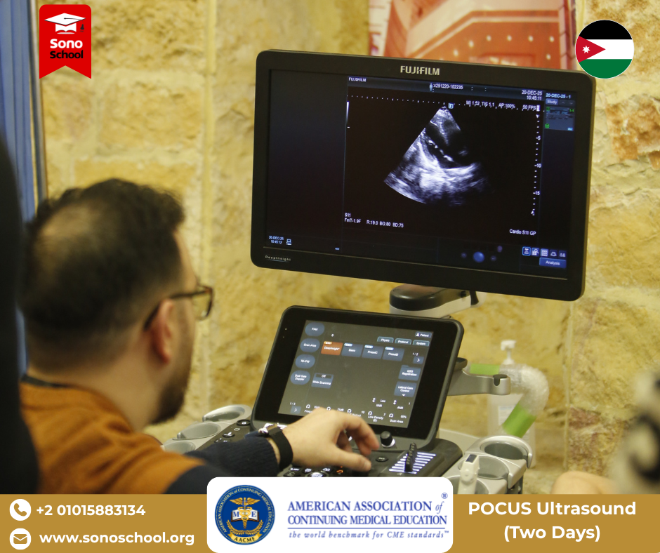
A cart-based ultrasound system may be controlled by a department, shared among trained users, or linked to established imaging workflows. A handheld ultrasound probe can be more personal, mobile, and immediately available. That convenience is valuable, but it also means the clinician needs a clear understanding of scope, limitations, and appropriate use.
This is where SonoSchool’s educational approach becomes important.
At SonoSchool, we do not view POCUS as device ownership alone. We view it as a clinical skill that requires structured learning, hands-on scanning, supervised practice, and continuous development.
The goal is not simply to help clinicians buy a handheld ultrasound device. The goal is to help them use ultrasound in a way that is clinically focused, technically competent, and integrated into real patient care.
Handheld POCUS Workflow Checklist
Use this checklist before introducing handheld ultrasound into your department, clinic, or personal clinical workflow.
| Workflow Area | Question to Answer | Why It Matters |
|---|---|---|
| Clinical scope | Which POCUS applications will we use? | Prevents random scanning and supports focused use. |
| Users | Who is allowed to scan? | Supports governance and patient safety. |
| Training | What training is required before independent use? | Device access does not equal competency. |
| Documentation | How will findings and images be recorded? | Supports clinical communication and QA. |
| Cleaning | How will the probe be cleaned between patients? | Maintains infection control workflow. |
| Escalation | When should formal imaging or expert review be requested? | Prevents over-reliance on limited bedside scans. |
A Practical Implementation Pathway
If you are introducing handheld POCUS into a clinical setting, start simple.
- Choose 2–3 priority applications.
For example: lung ultrasound, vascular access, and bladder assessment. - Train the core users first.
Start with clinicians who are most likely to use the device regularly. - Define minimum image requirements.
Clarify what a complete scan should include. - Create a documentation habit.
Decide how findings and limitations should be recorded. - Review cases regularly.
Use saved images and clips for quality improvement and learning. - Expand gradually.
Add cardiac, FAST/eFAST, procedures, or advanced applications as training develops.
This stepwise approach is safer than trying to use handheld ultrasound for every application from the beginning.
Final Takeaway
Handheld ultrasound can bring POCUS closer to the patient, but the device is only one part of the system.
To build handheld POCUS into real workflow, clinicians need clear clinical questions, defined applications, standard scanning approaches, documentation habits, cleaning processes, escalation rules, and structured training.
For many clinicians, the key shift is moving from:
“I own a probe.”
to:
“I know when to scan, how to scan, what the image means, how to document it, and when to escalate.”
That is the difference between owning handheld ultrasound and using POCUS well.
Build Handheld POCUS Into Your Clinical Workflow
SonoSchool supports clinicians and healthcare facilities with handheld ultrasound options, device guidance, specifications, availability information, and structured POCUS education pathways.
FAQ
How do I start using handheld POCUS in clinical workflow?
Start by defining a small number of focused applications, training the core users, standardizing image acquisition, deciding how findings will be documented, and setting clear escalation rules for formal imaging or expert review.
Is owning a handheld ultrasound enough to perform POCUS?
No. Owning a handheld ultrasound probe provides access to imaging, but POCUS competency requires training in image acquisition, interpretation, clinical integration, documentation, and understanding limitations.
Which POCUS applications should beginners start with?
The best starting applications depend on specialty and workflow. Common starting points include lung ultrasound, vascular access, bladder assessment, basic FAST/eFAST, and selected procedural guidance where training is available.
How should handheld ultrasound findings be documented?
Documentation should include the indication, views obtained, key findings, limitations, and how the scan influenced the clinical plan. Image or clip storage may also be required depending on institutional policy.
Can handheld POCUS be used for teaching?
Yes. Handheld ultrasound can be useful in training environments because of its portability and accessibility, but teaching should remain structured with supervision, defined objectives, and feedback.
When should handheld POCUS findings be escalated?
If findings are unclear, limited, inconsistent with the clinical picture, or outside the operator’s scope, clinicians should escalate to formal imaging, expert review, or senior clinical assessment.
Medical and Purchasing Disclaimer
This article is intended for educational and purchasing guidance only. Device selection and clinical use should follow local regulations, institutional policies, manufacturer documentation, and appropriate clinical training. Product-specific certifications, compatibility, warranty terms, pricing, and technical specifications should be verified directly before purchase.
What to explore next
This article is one step in a practical learning journey. Continue with related insights, hands-on workshops, or the full POCUS Fellowship pathway.
Recommended learning path
Choose the next step based on whether you want to read more, join a workshop, or explore structured fellowship training.
Ready to build real bedside ultrasound confidence?
Join structured POCUS courses and hands-on workshops designed to move clinicians from theory to confident bedside application.