
Critical Care Echo Explained: How to Master Bedside Cardiac Ultrasound in the ICU
Critical care echocardiography is an essential skill for physicians who manage critically ill patients in intensive care units (ICUs). When […]
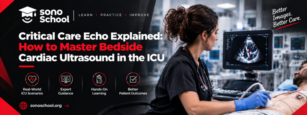
Critical care echocardiography is an essential skill for physicians who manage critically ill patients in intensive care units (ICUs). When a patient develops unexplained hypotension, shock, acute respiratory failure, or suspected cardiac dysfunction, bedside cardiac ultrasound can provide valuable information about cardiac structure and function in real time.
However, performing an ultrasound examination requires more than recognizing images on a screen. Physicians must understand cardiac anatomy, acquire appropriate views, interpret findings within the clinical context, and recognize when advanced imaging or specialist assessment is necessary.
A structured critical care echo course can help physicians develop these skills through systematic learning, practical scanning, and case-based clinical interpretation.
In this guide, you will learn what critical care echocardiography is, how it supports ICU decision-making, which views beginners should master, and how to choose the right training program to advance your clinical skills.
What Is Critical Care Echocardiography?
Critical care echocardiography is the use of ultrasound to assess cardiac structure and function in critically ill patients. It helps clinicians investigate cardiovascular problems at the bedside and integrate imaging findings into immediate clinical decisions.
Unlike a comprehensive echocardiographic examination performed by a cardiac imaging specialist, focused bedside echocardiography typically addresses specific clinical questions relevant to the patient’s condition.
Common applications include:
- Assessing global left ventricular (LV) systolic function.
- Evaluating right ventricular (RV) size and function.
- Identifying pericardial effusion and possible cardiac tamponade.
- Investigating potential cardiac causes of shock.
- Supporting the assessment of hemodynamic changes.
- Reassessing cardiac function during selected resuscitation scenarios.
Critical care echocardiography is particularly valuable when patient instability makes transport to an imaging department difficult.
According to the Society of Critical Care Medicine’s adult critical care ultrasonography guidelines, critical care ultrasound can support the evaluation and management of selected conditions, including septic shock, cardiogenic shock, and acute respiratory failure.
Why Is a Critical Care Echo Course Important for ICU Physicians?
Learning bedside echocardiography requires a combination of theoretical knowledge, practical scanning, and clinical reasoning. A structured training program helps physicians develop these skills systematically rather than relying exclusively on online videos or occasional bedside exposure.
1. Improve Bedside Cardiovascular Assessment
Echocardiography can help clinicians identify important cardiac abnormalities that may influence immediate management decisions. For example, reduced global LV function may support a cardiac contribution to shock, while significant RV abnormalities may prompt further investigation of right-sided pressure overload or other causes of RV dysfunction.
These findings must always be interpreted alongside the patient’s history, examination, laboratory results, and hemodynamic condition.
2. Support Shock Assessment
Shock can result from several mechanisms, including hypovolemia, cardiac dysfunction, distributive physiology, and obstructive causes.
Bedside cardiac ultrasound can help clinicians investigate these possibilities and determine which additional assessments may be required.
3. Develop Practical Scanning Confidence
Hands-on training helps physicians learn probe positioning, image optimization, anatomical orientation, and the acquisition of standard cardiac views.
4. Build a Foundation for Advanced Training
Once the fundamentals are established, physicians can progress toward advanced cardiac ultrasound, integrated hemodynamic assessment, and more complex critical care applications.
If you want to develop these skills systematically, explore SonoSchool’s ultrasound training programs and review the available options for emergency and critical care physicians.
How to Master Bedside Cardiac Ultrasound in the ICU: A Step-by-Step Guide
Step 1: Understand Basic Cardiac Ultrasound Principles
Before performing cardiac scans, learn the fundamentals of ultrasound physics and cardiac anatomy.
Focus on:
- Ultrasound probe types and their applications.
- Probe orientation and image markers.
- Depth, gain, and image optimization.
- Basic cardiac anatomy and chamber identification.
- Common artifacts and image-quality limitations.
- The difference between focused cardiac ultrasound and comprehensive echocardiography.
Understanding these principles makes it easier to recognize structures and obtain interpretable images.
Physicians who need to strengthen their general ultrasound skills can begin with the Basic POCUS Two Days Course at SonoSchool, which includes basic echocardiography alongside other bedside ultrasound applications.
Step 2: Master the Essential Echocardiographic Views
A systematic approach to image acquisition is fundamental to critical care echocardiography.
The following views are commonly used in focused bedside cardiac assessment.
| Cardiac view | What it helps assess |
| Parasternal long-axis (PLAX) | Cardiac chamber relationships, global LV appearance, and pericardial fluid |
| Parasternal short-axis (PSAX) | LV shape, selected regional abnormalities, and RV appearance |
| Apical four-chamber | Both ventricles, atria, and gross chamber-size relationships |
| Subcostal four-chamber | Cardiac structures and pericardial fluid, particularly when other windows are difficult |
| Inferior vena cava (IVC) view | Venous dimensions and respiratory variation as contextual hemodynamic information |
Each view provides different information. No single view is sufficient to answer every clinical question.
For example, the subcostal view may be helpful when a patient cannot tolerate the positioning required for standard parasternal or apical views. Conversely, poor acoustic windows may limit interpretation, making additional views or formal imaging necessary.
Practical tip: Learn the indications, limitations, and expected anatomical landmarks for each view before attempting complex interpretation.
Step 3: Assess Left Ventricular Function
Left ventricular function is an important component of critical care echocardiography.
In focused examinations, clinicians may assess global LV systolic function visually by examining myocardial movement and the apparent degree of ventricular contraction.
However, visual assessment is operator-dependent and should not be confused with a comprehensive quantitative evaluation of ejection fraction.
When assessing LV function, consider:
- Overall myocardial contraction.
- LV chamber size and geometry.
- Regional differences in wall motion, when assessable.
- Image quality and whether the available views are adequate.
- The patient’s clinical condition and relevant hemodynamic findings.
Reduced LV systolic function may contribute to a diagnosis of cardiogenic shock, but it does not independently establish the cause of shock or determine the appropriate treatment.
Step 4: Evaluate the Right Ventricle
Right ventricular assessment is particularly relevant in critically ill patients with respiratory failure, hemodynamic instability, or suspected right-sided cardiac dysfunction.
Important considerations include RV size relative to the LV, gross RV systolic function, septal configuration, and other findings relevant to the clinical question.
RV abnormalities can occur in several conditions, including pulmonary hypertension, acute pulmonary embolism, and complications of severe respiratory disease. However, RV enlargement or dysfunction is not specific to any single diagnosis.
A critical care echo course should teach clinicians how to recognize important patterns while understanding their limitations and the need for additional investigations when appropriate.
Step 5: Recognize Pericardial Effusion and Potential Tamponade
Pericardial effusion refers to fluid accumulation within the pericardial space. In selected patients, a significant effusion may impair cardiac filling and contribute to hemodynamic compromise.
Bedside echocardiography can help identify pericardial fluid and assess findings that may raise concern for cardiac tamponade.
Interpretation requires more than identifying fluid. Clinicians must consider the patient’s hemodynamic state, the size and distribution of the effusion, and relevant chamber-collapse findings.
Suspected tamponade in an unstable patient requires urgent clinical assessment and appropriate escalation. A limited bedside examination should not delay necessary emergency management.
Step 6: Integrate Echo Findings Into Shock Assessment
The greatest value of critical care echocardiography comes from combining imaging findings with the patient’s overall clinical presentation.
A structured assessment should include:
- Define the immediate clinical question.
- Obtain the appropriate cardiac views.
- Assess global LV and RV appearance.
- Look for significant pericardial fluid and other relevant findings.
- Integrate ultrasound observations with vital signs, examination findings, laboratory results, and available monitoring.
- Determine whether further imaging, specialist input, or urgent intervention is required.
- Repeat focused assessment when clinically indicated and appropriate.
For example, a hypotensive patient with reduced LV systolic function may require further assessment for cardiac dysfunction, while a patient with RV abnormalities and severe hypoxemia may need urgent investigation for potential pulmonary vascular disease.
These are examples of clinical reasoning, not standalone diagnostic rules. Treatment decisions must consider the complete clinical picture and relevant protocols.
For additional guidance, review the SCCM clinical guidelines on bedside cardiac ultrasonography, which discuss appropriate applications of cardiac ultrasound in critically ill patients.
How to Choose the Best Critical Care Echo Course
Selecting the right educational program is important for physicians who want to develop reliable bedside cardiac ultrasound skills.
Before you enroll in a critical care echo course, evaluate the following factors.
Structured curriculum: Choose a course covering cardiac anatomy, ultrasound physics, standard views, LV and RV assessment, pericardial effusion, and clinical interpretation.
Hands-on scanning: Practical sessions help learners develop probe-handling skills and improve image acquisition under instructor supervision.
Experienced instructors: Look for educators with relevant critical care, emergency medicine, or echocardiography experience.
Case-based teaching: Clinical scenarios help connect ultrasound findings with real ICU problems, including shock and respiratory failure.
Feedback and assessment: Training should include opportunities to review images, correct technical errors, and assess progress toward defined learning objectives.
Appropriate course level: Beginners should establish basic scanning skills before progressing to advanced hemodynamic interpretation.
SonoSchool lists a One Day Critical Care ECHO training option for rapid cardiac assessment in emergency and ICU settings. Visit SonoSchool’s official website or contact the training team to request the current curriculum, schedule, fees, eligibility requirements, and registration details.
Physicians seeking a broader and longer-term educational pathway can also explore the POCUS Fellowship in Acute Care Settings, which includes foundational learning followed by a specialized pathway in emergency medicine or critical care.
Frequently Asked Questions About Critical Care Echocardiography
1. What is a critical care echo course?
A critical care echo course teaches clinicians how to perform and interpret focused bedside cardiac ultrasound examinations in acute and intensive care settings. Depending on the program, it may cover cardiac views, LV and RV function, pericardial effusion, and the integration of findings into clinical assessment.
2. Who should take a critical care echocardiography course?
These courses may be appropriate for intensivists, emergency physicians, anesthesiologists, internal medicine physicians, and other clinicians involved in acute patient care. The ideal course level depends on previous ultrasound experience and professional responsibilities.
3. Can beginners learn critical care echo?
Yes, but beginners should first establish a foundation in ultrasound physics, cardiac anatomy, probe handling, and standard image acquisition. A focused introductory course may be suitable for some learners, while others may benefit from basic POCUS training before progressing to more advanced cardiac assessment.
4. How long does it take to learn bedside cardiac ultrasound?
The time required depends on the learner’s background, training quality, scanning opportunities, and assessment requirements. An introductory workshop can establish foundational knowledge, but reliable image acquisition and interpretation require ongoing supervised practice and competency assessment.
5. Is bedside echocardiography enough to diagnose every cardiac condition?
No. Focused bedside echocardiography answers specific clinical questions and may not provide all the information available from a comprehensive echocardiographic study. Inconclusive findings or suspected complex cardiac disease may require formal imaging and specialist assessment.
6. What is the difference between POCUS and critical care echocardiography?
POCUS is a broad term covering focused ultrasound examinations across multiple organ systems. Critical care echocardiography specifically focuses on cardiac structure and function in critically ill patients, often as part of a wider assessment of shock, respiratory failure, and hemodynamic instability.
7. Where can I enroll in a critical care echo course?
You can explore SonoSchool’s training programs and contact the team for Critical Care ECHO course information. Request details about upcoming dates, practical training, fees, course requirements, and certification policies before registering.
Advance Your ICU Ultrasound Skills With Structured Training
Critical care echocardiography can provide valuable information when evaluating cardiac function and hemodynamic instability in critically ill patients. Developing this skill requires a structured learning pathway that combines anatomical knowledge, systematic image acquisition, careful interpretation, and supervised practice.
For physicians seeking to improve their bedside assessment skills, the right critical care echo course can provide an important starting point for continued professional development.
Explore SonoSchool’s ultrasound training options, compare the available educational pathways, and contact the team to learn more about Critical Care ECHO training and upcoming course dates.
Build your skills progressively, seek expert feedback, and always interpret ultrasound findings within the wider clinical context. Competency-based training and appropriate clinical supervision remain essential for safe and effective bedside echocardiography.
Recommended learning path
Choose the next step based on whether you want to read more, join a workshop, or explore structured fellowship training.


Ready to build real bedside ultrasound confidence?
Join structured POCUS courses and hands-on workshops designed to move clinicians from theory to confident bedside application.