
5 Clinical Situations Where Handheld Ultrasound Can Change Your Next Decision
Clinical Need In acute care, the problem is not always a lack of clinical knowledge. Often, the problem is distance. The ultrasound machine is across the department. The…
Clinical Need
In acute care, the problem is not always a lack of clinical knowledge. Often, the problem is distance.
The ultrasound machine is across the department. The patient is unstable. The procedure needs to start. The ward call is urgent. The clinic is busy. The decision cannot wait for a perfect imaging workflow.
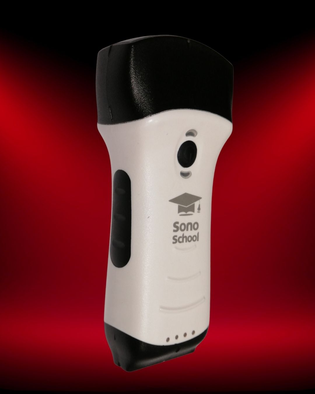
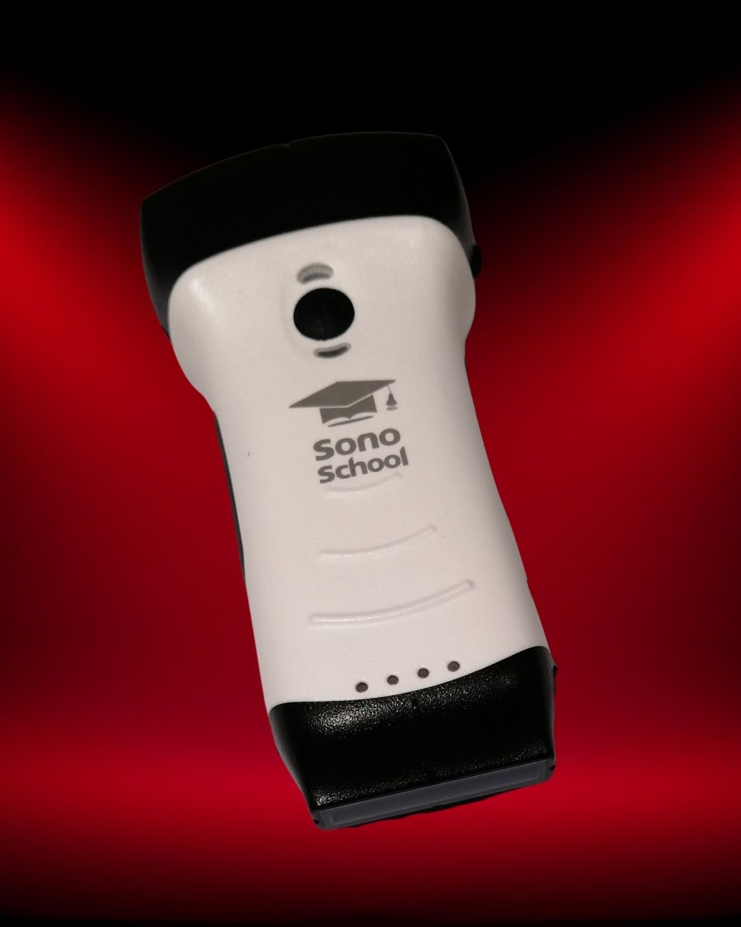
This is where handheld ultrasound becomes clinically interesting. A handheld ultrasound device does not replace clinical judgment, formal imaging, or comprehensive ultrasound systems. It also does not remove the need for proper POCUS training. But when used by trained clinicians within an appropriate scope, handheld ultrasound can bring focused imaging closer to the bedside and help answer practical clinical questions in real time.
The key word is focused. Point-of-care ultrasound is not about scanning everything. It is about asking a specific clinical question and using ultrasound findings alongside history, examination, vital signs, labs, and the wider clinical picture.
1. Acute Dyspnea: Is This Lung, Heart, Fluid, or Something Else?
A patient arrives short of breath.
The differential is broad: pulmonary edema, pneumonia, pleural effusion, pneumothorax, COPD/asthma exacerbation, pulmonary embolism, heart failure, sepsis, or mixed pathology. Clinical examination is essential, but it can be limited by noise, body habitus, mechanical ventilation, or overlapping signs.
A handheld ultrasound probe may help the clinician rapidly assess several key bedside findings:
- Pleural sliding
- B-lines
- A-lines
- Pleural effusion
- Consolidation patterns
- Basic cardiac function
- Pericardial effusion
- IVC appearance, when clinically relevant
This does not mean handheld ultrasound gives a complete diagnosis by itself. It means it can help refine the next decision.
How it may influence the next decision
- Diffuse bilateral B-lines with reduced cardiac function may support a cardiogenic pulmonary edema pathway.
- A large pleural effusion may change drainage planning or imaging escalation.
- Absent lung sliding in the right clinical context may raise concern for pneumothorax.
- Focal lung findings may make pneumonia or localized pathology more likely.
What to check before relying on handheld ultrasound in dyspnea
A handheld ultrasound device used for respiratory assessment should provide enough image quality for lung artifacts, pleural line assessment, effusions, and basic cardiac views where required. For clinicians working in ER, ICU, internal medicine, anesthesia, or acute clinics, lung and focused cardiac scanning should be tested before purchase.
2. Shock and Hypotension: What Direction Should Resuscitation Take?
A hypotensive patient needs decisions quickly.
Is the likely problem hypovolemia, cardiac dysfunction, obstructive physiology, bleeding, sepsis, or a mixed picture? No single scan answers every question, and ultrasound findings should never be interpreted in isolation. But focused bedside ultrasound can help guide the direction of resuscitation.
In shock, clinicians may use POCUS to assess:
- Gross LV function
- RV size and strain pattern, when visible
- Pericardial effusion
- IVC appearance and respiratory variation, with caution
- Free fluid in trauma or suspected bleeding
- Lung findings suggesting pulmonary edema or fluid tolerance concerns
- Abdominal aorta in selected patients
- Volume status clues, interpreted alongside the full clinical picture
A handheld ultrasound device can be useful here because the patient may not be easy to move. If the device is immediately available, the clinician can repeat focused scans during resuscitation rather than treating ultrasound as a one-time event.
Clinical framing: The handheld ultrasound is not replacing the resuscitation team, senior review, laboratory data, or formal imaging. It is helping the clinician decide what to do next, faster and with more context.
How handheld ultrasound may influence the next decision
- Whether to prioritize fluids, vasopressors, or inotropes
- Whether cardiac dysfunction needs urgent consideration
- Whether obstructive causes need escalation
- Whether pulmonary edema limits further fluid administration
- Whether free fluid changes the urgency of surgical or imaging escalation
3. Trauma or Acute Abdominal Concern: Is There a Bedside Clue That Changes Urgency?
In trauma, abdominal pain, collapse, or suspected internal bleeding, time matters.
Focused ultrasound can help clinicians look for free fluid in selected clinical contexts. In trauma workflows, FAST/eFAST is commonly used to assess for free fluid and thoracic findings as part of bedside evaluation.
For handheld ultrasound, the question is not, “Can it replace full imaging?”
It cannot.
The better question is:
Can it provide an immediate bedside clue that changes urgency, escalation, or monitoring?
Examples include:
- A trauma patient with hypotension and suspected intra-abdominal bleeding
- A patient with abdominal pain and hemodynamic instability
- A bedside reassessment when transport to imaging is delayed
- A resource-limited setting where immediate formal imaging is unavailable
- A patient requiring serial focused reassessment
Practical limitation
Handheld ultrasound should not create false confidence. Negative bedside findings do not automatically exclude significant pathology. Clinical context, operator skill, patient factors, and image quality all matter. When the clinical picture remains concerning, escalation should not be delayed because of a limited handheld scan.
4. Vascular Access and Procedures: Can You Make the Next Attempt Safer and More Deliberate?
Few situations show the practical value of bedside ultrasound as clearly as vascular access and procedural guidance.
When vessels are small, deep, collapsed, scarred, or difficult to palpate, ultrasound can help clinicians identify anatomy before puncture and guide needle placement dynamically.
For a handheld ultrasound device, procedural use may include:
- Peripheral IV access
- Central venous access support
- Arterial line placement support
- Abscess evaluation and drainage planning
- Pleural procedure planning
- Nerve block guidance, where within scope
- Soft tissue assessment
- Foreign body localization in selected cases
This is one of the strongest practical arguments for having ultrasound close to the clinician. If the machine is far away, unavailable, or difficult to move, ultrasound guidance may be skipped even when it could help. A handheld ultrasound probe can lower that access barrier.
What matters most for procedural use
For procedural applications, image quality is only one part of the decision.
- Near-field resolution
- Needle visualization
- Screen size and brightness
- Latency
- Probe ergonomics
- Sterile cover compatibility
- Battery life
- Ease of cleaning
- Whether the app allows fast adjustments during the procedure
For vascular access and nerve block workflows, a linear probe configuration is usually important. For pleural or deeper procedural planning, other probe types may be needed.
Training point: Seeing the vessel is not the same as safely guiding the needle. Clinicians need supervised practice in probe handling, needle alignment, hand-eye coordination, and recognition of complications.
5. Reassessment at the Bedside: Has the Patient Changed Since the Last Review?
One of the most useful roles of handheld ultrasound is not the first scan.
It is the repeat scan.
Many clinical decisions depend on change over time:
- Is pulmonary edema worsening after fluids?
- Is pleural effusion increasing?
- Is bladder volume contributing to discomfort or retention?
- Has lung aeration changed?
- Is cardiac function grossly different from the earlier assessment?
- Is the patient tolerating ongoing resuscitation?
- Does the procedure site need reassessment?
A cart-based ultrasound system may still be preferred for comprehensive imaging. But handheld ultrasound can make focused reassessment easier because the device can remain close to the clinical team.
The clinical value is not only portability. It is repeatability.
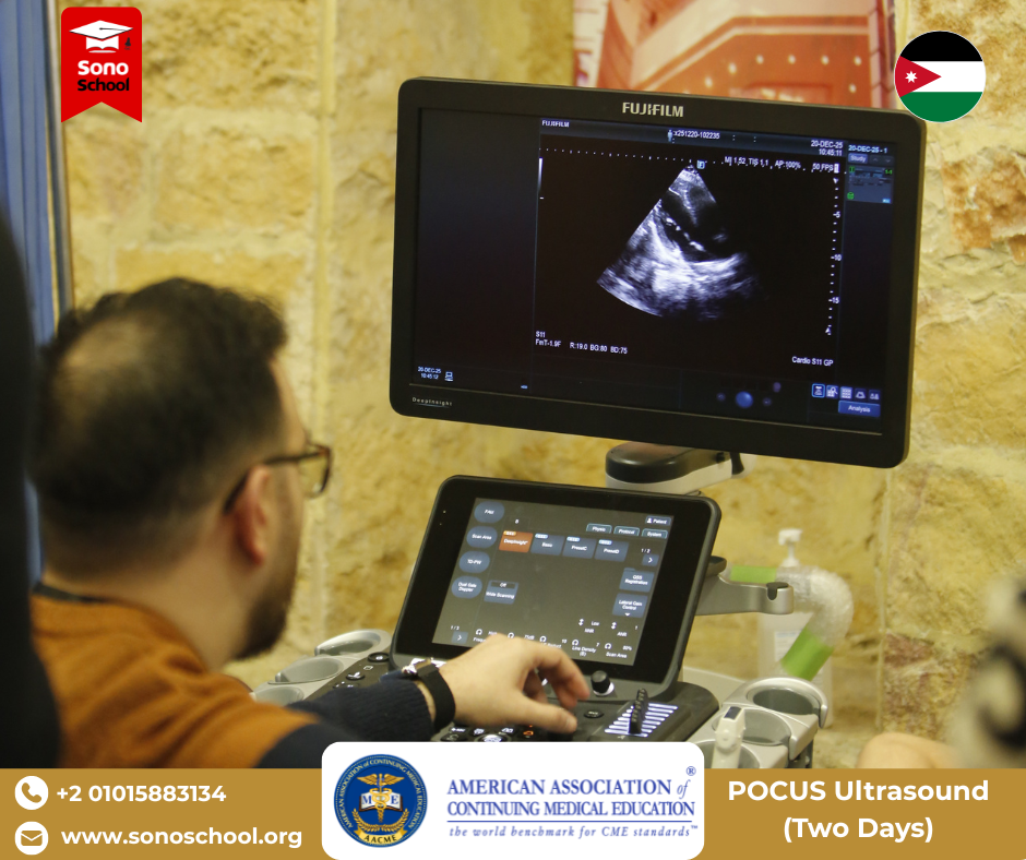
This is especially relevant in ICU, emergency departments, step-down units, wards, outpatient clinics, home-care or remote-care models, training environments, and resource-limited settings.
The risk of repeat scanning
Repeated scanning should not mean overconfidence or overinterpretation. A focused bedside ultrasound exam should answer a defined question. If the finding is unclear, limited, or clinically inconsistent, formal imaging or expert review remains necessary.
What Handheld Ultrasound Should Not Be Expected to Do
A balanced buying decision requires understanding limitations.
A handheld ultrasound device should not be expected to:
- Replace comprehensive radiology-performed ultrasound
- Replace echocardiography when detailed measurements are needed
- Make clinicians competent without training
- Provide reliable answers in every patient
- Overcome poor scanning technique
- Solve documentation or governance issues by itself
- Replace clinical judgment
- Replace escalation when the patient is unstable
Handheld ultrasound is most useful when the question is focused, the operator is trained, the device matches the application, and the clinical team understands the limitations.
From Device Access to Clinical Confidence
The biggest mistake clinicians can make is thinking of handheld ultrasound only as a product.
The better way to think about it is as a clinical workflow.
A point-of-care ultrasound device becomes valuable when four elements come together:
- The right clinical question
The scan should be performed for a defined reason. - The right probe and image quality
The device should match the intended application. - The right workflow
Images, clips, documentation, cleaning, storage, and export need to fit the clinical environment. - The right training
Clinicians need structured education, supervised practice, and ongoing feedback.
This is where SonoSchool’s role goes beyond the device itself.
At SonoSchool, we believe POCUS is not simply about owning an ultrasound probe. It is about developing the ability to acquire images, interpret them correctly, and integrate findings into clinical decisions with confidence.
A handheld ultrasound device can bring imaging closer to the patient. Training is what helps clinicians use that access responsibly.
Clinical Situations Summary
| Clinical Situation | How Handheld Ultrasound May Help | Key Limitation |
|---|---|---|
| Acute dyspnea | Supports focused lung and cardiac assessment | Findings must match clinical context |
| Shock and hypotension | Helps guide resuscitation direction | Does not replace senior review or full workup |
| Trauma or acute abdomen | May provide bedside clues about free fluid or urgency | Negative findings do not exclude pathology |
| Vascular access and procedures | Supports anatomy identification and dynamic guidance | Requires procedural ultrasound skill |
| Bedside reassessment | Allows focused repeat scanning over time | Repeated scanning should remain question-driven |
Choosing a Handheld Ultrasound for These Situations
If you are considering a handheld ultrasound probe for emergency, critical care, anesthesia, internal medicine, surgery, general practice, or clinic use, evaluate the device against your most common clinical decisions.
Ask yourself:
- Which five clinical situations would I use it for most?
- Which probe configuration do those situations require?
- Do I need lung, cardiac, abdominal, vascular, or procedural imaging?
- Is the device compatible with my phone, tablet, or clinical hardware?
- Can I save and export images appropriately?
- Is DICOM required in my facility?
- What support is available in my country?
- What training pathway is available for me or my team?
A handheld ultrasound device should not just be easy to carry. It should be clinically useful, supportable, and teachable.
Explore Handheld Ultrasound Options Available Through SonoSchool
For clinicians and healthcare facilities considering handheld ultrasound, SonoSchool can support the decision-making process with device guidance, specifications, availability information, and POCUS education pathways.
FAQ
What clinical situations are best suited for handheld ultrasound?
Handheld ultrasound is commonly useful for focused bedside questions such as dyspnea assessment, shock evaluation, vascular access, procedural guidance, trauma screening, bladder assessment, lung ultrasound, and reassessment. Suitability depends on the device, operator skill, and clinical context.
Can handheld ultrasound be used in emergency and ICU settings?
Yes, handheld ultrasound may be useful in emergency and ICU settings when used by trained clinicians within an appropriate workflow. It can support focused bedside assessment, but it should not replace formal imaging or comprehensive evaluation when these are required.
Is handheld ultrasound useful for vascular access?
Handheld ultrasound can be useful for vascular access if the image quality, probe type, and screen workflow are appropriate. Clinicians still need training in needle visualization, dynamic guidance, and procedural safety.
Does handheld ultrasound replace cart-based ultrasound?
No. Handheld ultrasound does not replace every cart-based ultrasound system. It is best viewed as a portable bedside tool for focused questions. Cart-based systems may still be preferred for advanced imaging, formal studies, and comprehensive examinations.
Do clinicians need training before using handheld ultrasound?
Yes. A handheld ultrasound probe only provides access to imaging. Competency requires training in image acquisition, interpretation, protocols, documentation, and clinical integration.
What should I check before buying a handheld ultrasound device?
Check image quality, probe configuration, compatibility, workflow, storage/export options, DICOM requirements, regulatory documentation, warranty, regional support, total cost, and training availability.
Medical and Purchasing Disclaimer
This article is intended for educational and purchasing guidance only. Device selection and clinical use should follow local regulations, institutional policies, manufacturer documentation, and appropriate clinical training. Product-specific certifications, compatibility, warranty terms, pricing, and technical specifications should be verified directly before purchase.
What to explore next
This article is one step in a practical learning journey. Continue with related insights, hands-on workshops, or the full POCUS Fellowship pathway.
Recommended learning path
Choose the next step based on whether you want to read more, join a workshop, or explore structured fellowship training.
Ready to build real bedside ultrasound confidence?
Join structured POCUS courses and hands-on workshops designed to move clinicians from theory to confident bedside application.